
When a tooth is lost, the most obvious change is the gap that appears in the dental arch. However, a more subtle process also takes place beneath the gums: the jawbone at the missing-tooth site begins to change and gradually lose volume.
This process usually does not cause pain, so many people do not notice it. Years after tooth loss, when they consider dental implant treatment, some patients only then discover that the jawbone has lost height or width, making the restorative plan more complex.
So how does the jawbone resorb after tooth loss? Why does the risk of insufficient bone increase the longer a tooth is missing? And what can be done to help limit this condition?
The alveolar bone is the part of the jawbone that surrounds and supports the tooth roots. While a tooth is present, forces generated during chewing are transmitted through the root to the surrounding bone.

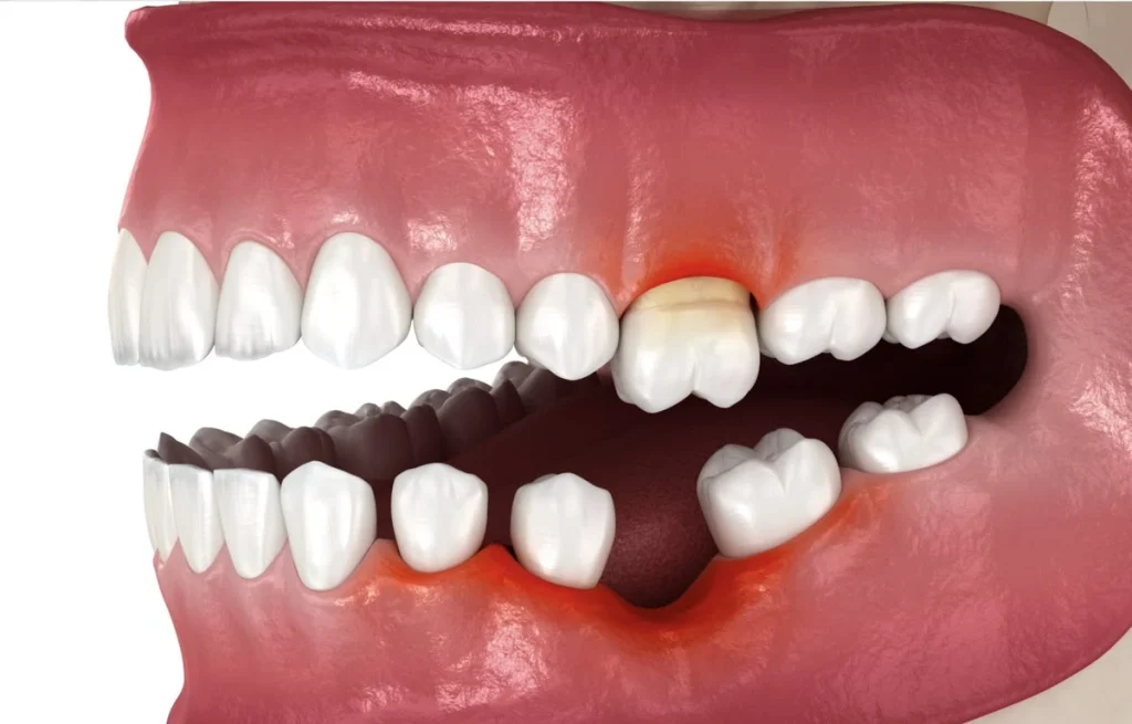
What is jawbone loss after tooth loss
After a tooth is lost, the biological environment in the area changes. The socket undergoes healing and remodeling, while the dimensions of the alveolar ridge tend to decrease in both width and height. This is considered part of the normal physiological remodeling process after tooth extraction.
Simply put:
Tooth present → the jawbone is maintained in a functional load-bearing environment.
Tooth lost → the socket heals and the alveolar ridge begins to remodel → the bone dimensions at the missing-tooth site may gradually decrease.
It is important to note that this process occurs beneath the gums, so patients usually cannot see it immediately with the naked eye.
Bone loss does not happen suddenly in a single day. It is a remodeling process that develops over time and is often more pronounced during the early period after tooth loss.
After a tooth is removed from the jawbone, a blood clot forms in the socket and the healing process begins.
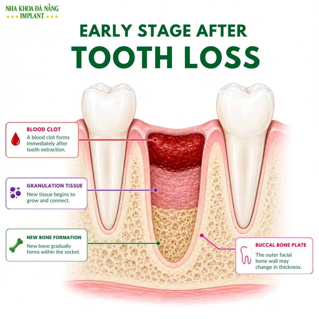
Early Stage After Tooth Loss
The body gradually forms new tissue and new bone inside the socket. However, alongside the healing process, the walls of the tooth socket also begin to remodel.
Therefore, the socket being “filled in” does not mean that the shape and dimensions of the jawbone will completely return to what they were before extraction.
This is the most notable stage.
Studies on post-extraction healing show that most dimensional changes of the alveolar ridge occur during the first few months, particularly in ridge width. The facial/buccal bone wall generally tends to change more than the inner wall.
One systematic review reported an average reduction in alveolar ridge width of approximately 3.87 mm, while the average reduction in facial bone height was about 1.67 mm. However, the actual degree of change can vary considerably from person to person.
This helps explain why a recently lost tooth site may still have enough bone for restorative planning, while after a long period of time the bone conditions may no longer be as favorable as they were initially.
After the initial period of rapid change, bone remodeling may continue over time.
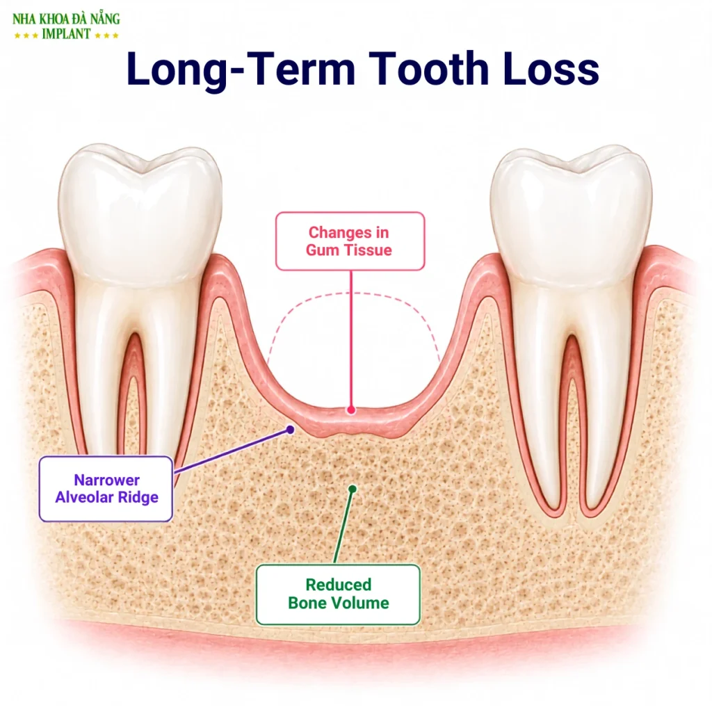
Jawbone Resorption After Long-Term Tooth Loss
If the missing tooth is not appropriately restored, the edentulous area may increasingly show:
When a tooth has been missing for many years, the degree of bone loss varies from person to person depending on the tooth position, previous periodontal condition, cause and duration of tooth loss, bone anatomy, general health, and many other factors.
Therefore, the question “How many years has my tooth been missing?” alone cannot determine whether there is still enough bone for dental implant placement.
Bone is living tissue that is continuously remodeled.
When a tooth is present, the root and periodontal tissues help transmit chewing forces to the surrounding bone. After tooth loss, the structure and mechanical environment of that area change. Based on the principle of bone adaptation to loading, changes in mechanical stimulation are among the factors associated with reduced alveolar ridge dimensions after tooth loss.
In addition, tooth extraction is followed by a natural biological remodeling process of the socket walls themselves.
Therefore, bone loss after tooth loss is not simply “the bone disappearing because you no longer chew there.” It results from healing, bone remodeling, and changes in the functional environment after the tooth is no longer present.
In the early stages, jawbone loss often progresses quietly and does not necessarily cause pain.
When the condition progresses over a long period, patients may notice signs such as:
The gums at the missing-tooth site appear sunken
As the underlying bone loses volume, the gum contour may change and the missing-tooth area can appear more depressed than before.
Changes in the missing-tooth space
Adjacent teeth may gradually tilt into the gap, while the opposing tooth may over-erupt toward the missing-tooth space.
Difficulty chewing
When several teeth are missing, patients may shift chewing to the side where more teeth remain, reducing chewing efficiency and causing uneven distribution of chewing forces.
Facial changes in cases of multiple missing teeth
When many teeth or a full arch of teeth have been missing for a long time, reduced support for the lips, cheeks, and lower third of the face may make the facial appearance look older.
However, the degree of bone loss cannot be accurately assessed by visual inspection alone.
Not every long-standing missing-tooth case is unsuitable for dental implants.
However, when the alveolar ridge has significantly decreased in width or height, implant placement may become more complex.
Dental implants require suitable bone conditions so the dentist can safely select the position, size, and angulation of the implant. Therefore, when bone volume is insufficient, the dentist may need to consider additional procedures depending on the individual case, such as:
Professional reviews also note that reductions in alveolar ridge height and width after tooth loss can become an obstacle to placing implants in an optimal position.
For this reason, early examination and restorative planning generally provide dentists with more favorable treatment options than waiting until substantial bone changes have already occurred.
This is a common point of misunderstanding.
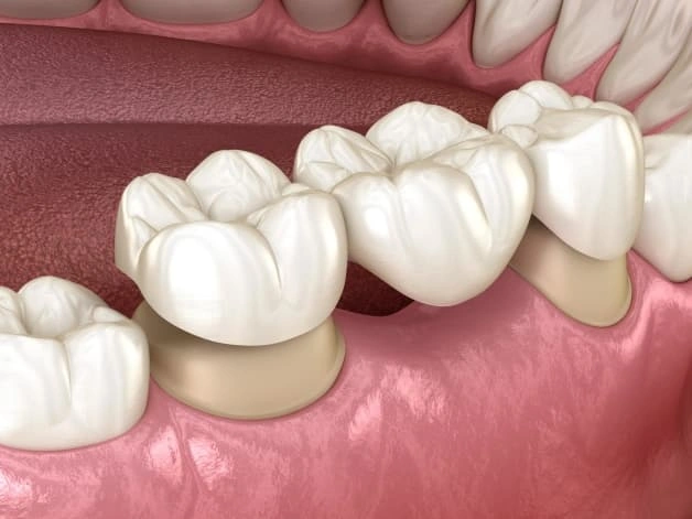
Dental Bridge
Dental bridge
A dental bridge can restore the visible tooth structure above the missing-tooth space and improve chewing function. However, the missing tooth no longer has a root within the corresponding bone area.
In addition, this method often requires the adjacent teeth to be used as abutments.
Removable denture
A removable denture can restore multiple missing teeth at a lower initial cost, but chewing forces are transmitted mainly through the denture base and soft tissues.
Therefore, both methods have their own restorative roles, but neither recreates a tooth root positioned directly within the bone in the same way as a dental implant.
A dental implant is a fixture placed into the jawbone to serve as a foundation for the dental restoration above it.
Compared with leaving the missing-tooth space untreated, dental implants offer several notable advantages:
However, it is important to understand that dental implants cannot guarantee that “bone will never resorb.” Bone around an implant is still living tissue and is influenced by oral hygiene, loading, periodontal status, smoking, systemic conditions, and many other factors.
Therefore, long-term outcomes depend greatly on the initial treatment plan as well as proper care and regular follow-up after treatment.
There is no single timeline that applies to every patient.
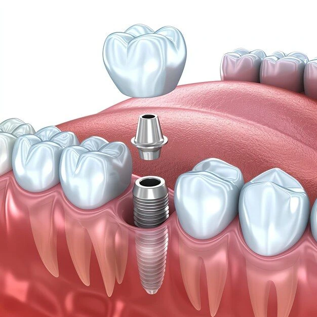
Dental Implant Illustration
Depending on the specific condition and the extent of jawbone loss, the dentist may consider:
Studies show that ridge-preservation techniques after extraction may help limit the reduction in bone dimensions and reduce the progression of jawbone loss compared with allowing the socket to heal naturally, although they cannot completely prevent physiological bone remodeling..
The most important point is this: if you have lost a tooth, do not wait until you are in pain or have difficulty chewing before seeking an examination.
An early consultation allows the dentist to assess your current bone condition, determine the extent of jawbone loss, and choose the most appropriate time and treatment approach for restoring the missing tooth.
In some cases, visual examination of the gums or a conventional two-dimensional X-ray may not be sufficient to fully assess jawbone loss and the bone structure required for implant planning.
The dentist needs to combine a clinical examination with appropriate diagnostic imaging to assess:
In implant dentistry, Cone Beam Computed Tomography (CBCT) allows three-dimensional assessment of bone structures and supports the dentist in planning the appropriate implant position and angulation.
Therefore, even if you have been missing a tooth for 5 years, 10 years, or longer, you should not assume that severe jawbone loss means you have “no bone left” or that dental implant treatment is no longer possible. A proper examination and CBCT assessment are necessary to determine your current bone condition and the most suitable treatment plan.
Several important principles include:
Seek an early examination after tooth loss or extraction
The dentist can assess the condition of the socket and plan the restorative direction from the beginning.
Consider socket preservation when indicated
In some cases, the dentist may perform ridge-preservation techniques after extraction to reduce changes in bone height and width.
Restore missing teeth at an appropriate time
A missing-tooth space should not be left untreated for many years simply because it does not yet feel inconvenient.
Treat periodontal disease if present
Periodontitis can also cause loss of the bone that supports the teeth. Good periodontal control is very important before and after implant restoration.
Maintain good oral hygiene and attend regular follow-up visits
Even after implant restoration, patients still need proper oral hygiene and periodic examinations to maintain the health of the tissues around the implant.
In many cases, yes, but the condition must be individually assessed.
Modern implant dentistry offers a range of treatment options for patients with long-standing tooth loss, insufficient bone, or complex bone loss.
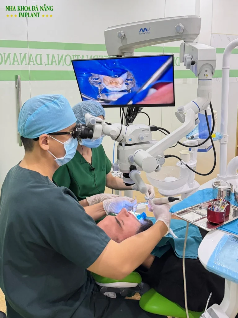
Da Nang Implant Dental Clinic
Depending on the patient, the dentist may consider:
No two cases of bone loss are exactly the same. Therefore, one patient’s treatment plan should not be applied mechanically to another patient.
An accurate diagnosis is the most important step before making a treatment decision.
For patients with long-standing tooth loss, the key question is not only “Can an implant be placed?” It is also necessary to determine:
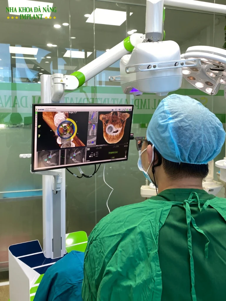
Jawbone Assessment at Da Nang Implant Dental Clinic
How much bone remains? Where should the implant be placed? Which angulation is safe? Is bone grafting required? And which treatment option best matches the patient’s actual condition?
At Da Nang Implant Dental Clinic, implant treatment is based on a detailed examination and individualized planning for each patient, including:
If you have been missing teeth for many years and are concerned that your jawbone has resorbed, do not assume too quickly that dental implant treatment is no longer possible.
Have your bone condition assessed first. Based on actual diagnostic data, the dentist can determine the most appropriate treatment plan and help avoid unnecessary interventions.
—————–
Address: 420 2/9 Street, Hoa Cuong Ward, Da Nang City
Address: 420 2/9 Street, Hoa Cuong Ward, Da Nang City
Hotline: (+84) 899 412 412
cskh@nhakhoaimplantdanang.com

Ngày: 19/04/2025

Ngày: 26/10/2023

Ngày: 14/10/2026
Ngày: 13/10/2026
Ngày: 08/10/2026
Ngày: 05/10/2026